
Neural Signal Pathway
Diagram showing neural signal transmission from sensory input through thalamus to cortex
Neuroanatomy
Draft neurology visuals for lesion localization, neural pathways, cranial nerve syndromes, spinal cord findings, EEG placement, and neurodiagnostic teaching. This page is narrower and more clinical than the broader nervous-system overview page.
Free to try ·
Your diagram will appear here
Describe what you need and click Generate
Professional neuroscience diagrams generated with AI
Diagram showing neural signal transmission from sensory input through thalamus to cortex
Cross-sectional view showing the six layers of the cerebral cortex with cell types
Color-coded brain map showing primary functional areas and their roles
A high-information-density teaching figure with a main view, detail insets, legend, and review-ready labels for spinal cord pathways.
A high-information-density teaching figure with a main view, detail insets, legend, and review-ready labels for neuromuscular junction.
A high-information-density teaching figure with a main view, detail insets, legend, and review-ready labels for autonomic reflex arc.
Neurology diagrams work best when the question is clinical: where a lesion localizes, how a pathway explains a deficit, or how anatomy supports a neurologic exam, syndrome, or diagnostic workflow.
This page is useful for neurologists, neuroscience educators, trainees, and presenters who need pathway logic and clinical relevance in the same figure.
Include the pathway, the exam finding, or the syndrome directly in the prompt. If the need is mainly brain structure, a brain-anatomy page is often cleaner. If the goal is a full CNS/PNS overview, the nervous-system page is usually broader and more appropriate.
These figures are common in bedside teaching, board review, clinic explanation, case conferences, and talks where lesion logic has to be understood quickly.
Generate professional neuroscience illustrations for teaching, clinical practice, and research presentations.
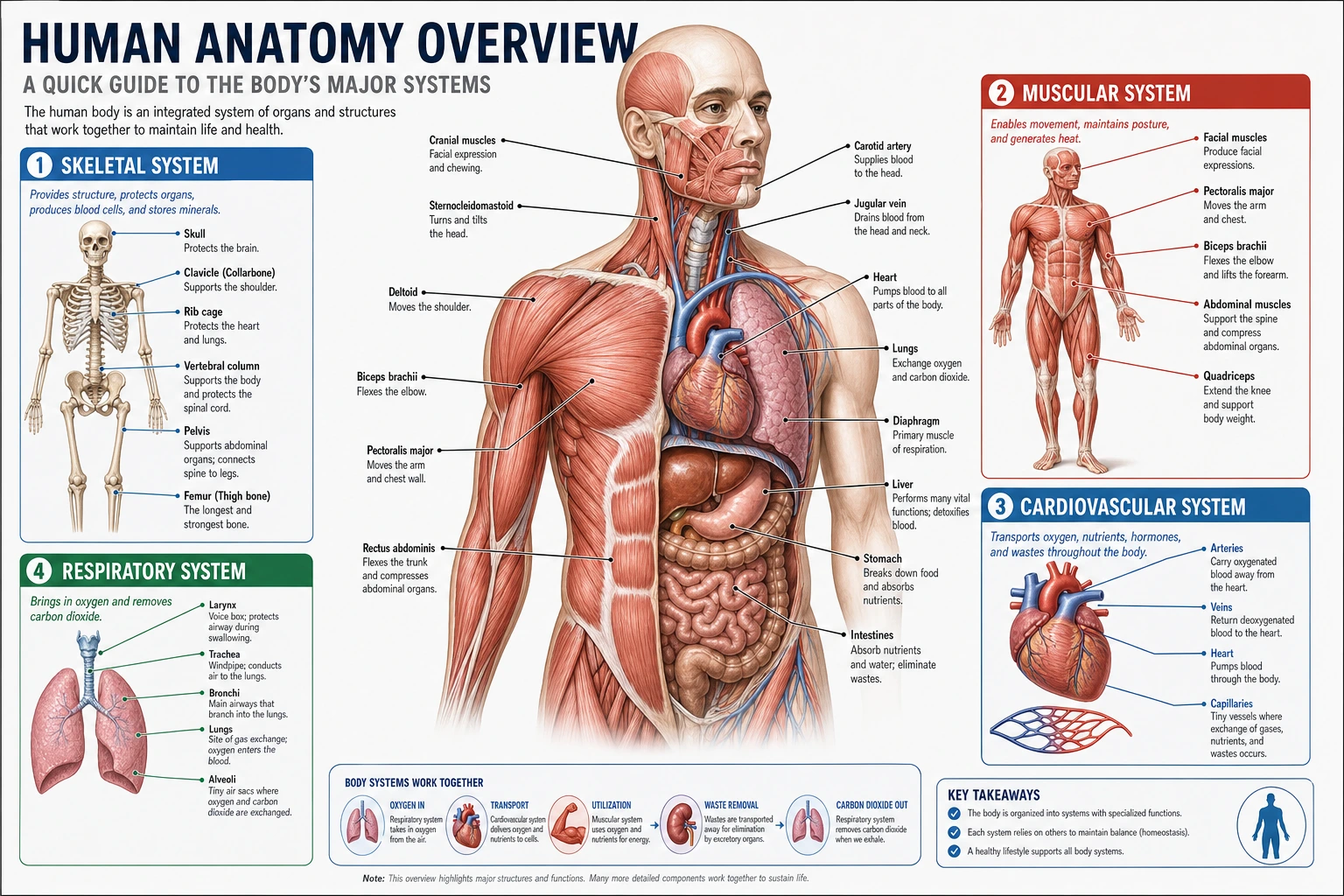 Anatomy
General-purpose anatomical diagram generator for all body systems
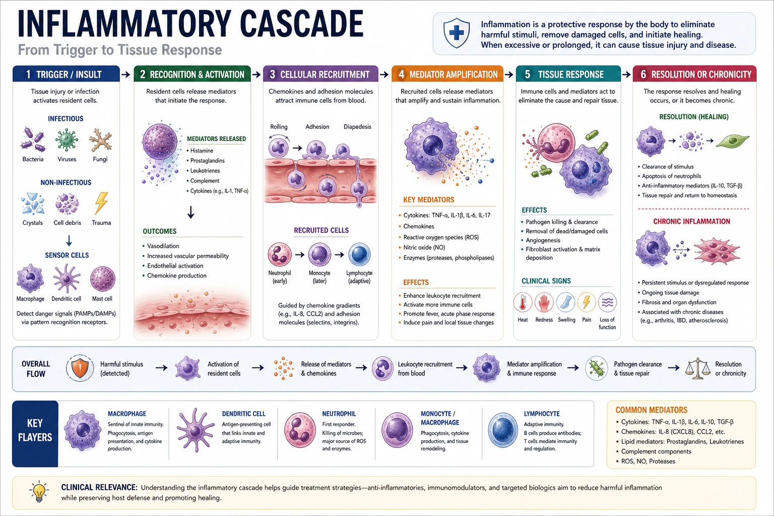 Clinical
Create clinical diagrams for pathways, mechanisms, and processes
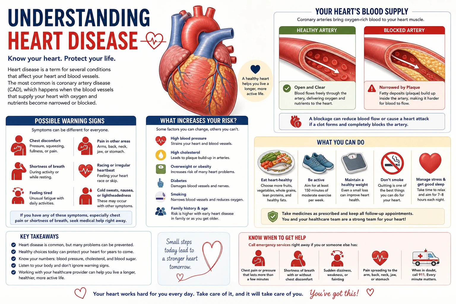 Education
Create simplified brain and nervous system visuals for patient handouts