
Eye globe overview
A labeled draft showing cornea, lens, retina, vitreous, and optic-nerve relationship in one view.
Eye Anatomy
Draft ophthalmology visuals for eye anatomy, retinal layers, aqueous flow, lens structure, and clinic explanation. Define the eye region, the view, and the audience before you generate.
Free to try ·
Your diagram will appear here
Describe what you need and click Generate
Example eye and retinal drafts for teaching, clinic explanation, and study materials.
A labeled draft showing cornea, lens, retina, vitreous, and optic-nerve relationship in one view.
A layer-focused draft that organizes retinal anatomy for study and clinic explanation.
A process-meets-structure draft that clarifies the main fluid path and drainage points.
A simplified draft that helps explain lens position and change across common teaching scenarios.
A labeled draft that groups the eye-movement muscles and their main direction of action.
A teaching draft that links retina, optic nerve, chiasm, tract, and cortical destination at a high level.
Use this page when the request is mainly about eye structure, retinal organization, fluid-flow teaching, or simplified ophthalmology explanation rather than a full surgical or disease workflow.
Strong ophthalmology prompts define the eye region, the perspective, and whether the emphasis should sit on anatomy, flow, or a comparative explanation.
This page helps with first-pass eye-anatomy explanation, but final review should still confirm structure naming, flow logic, and any patient-facing simplification.
Use another page when the visual should focus more on patient handouts, brain-pathway context, or clinician-facing disease mechanisms rather than eye anatomy itself.
Describe the eye region, the labels, and the audience, then review the output against your trusted source before reuse.
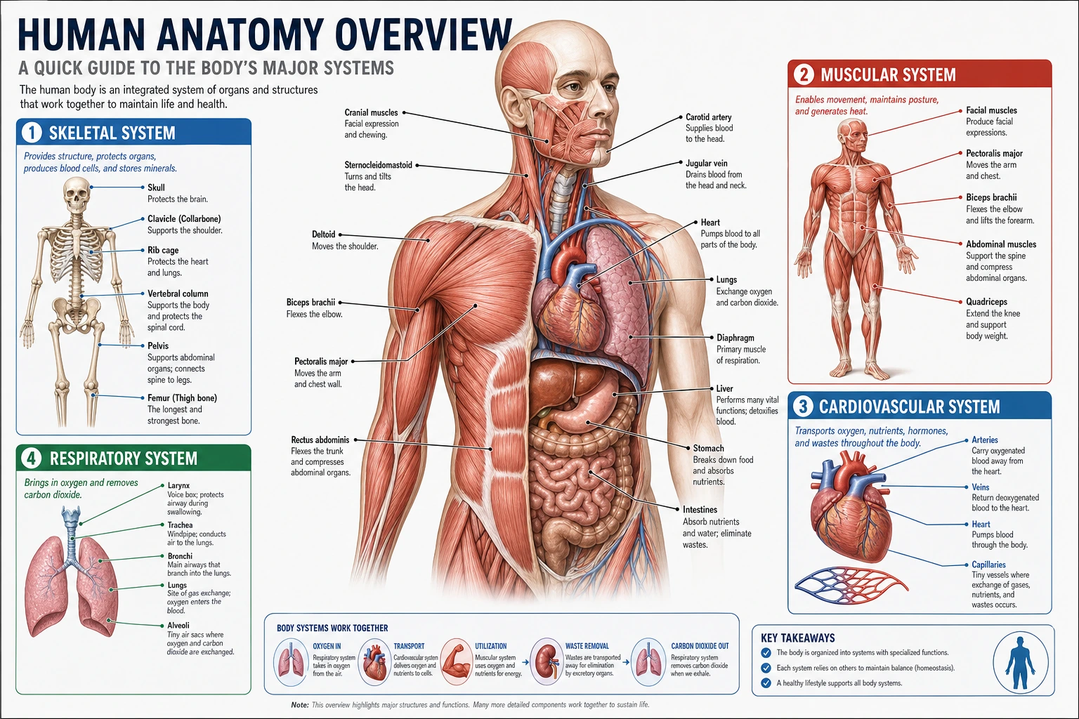 Anatomy
Use a broader anatomy page for head-and-neck or mixed-structure teaching visuals beyond the eye alone.
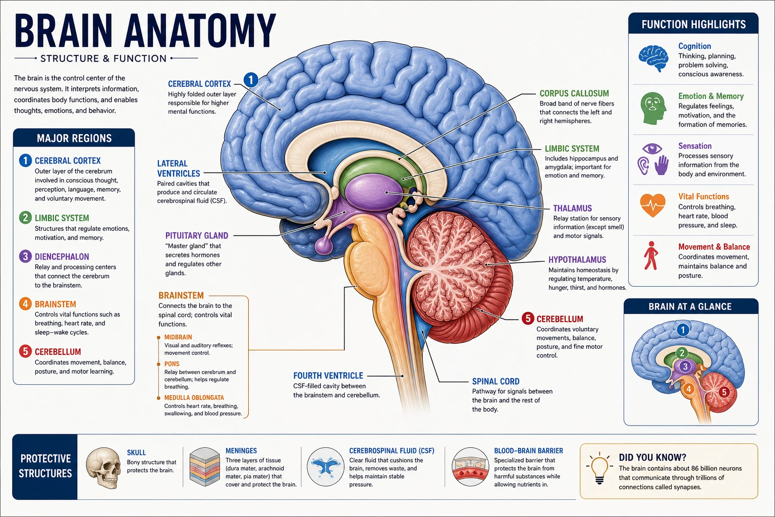 Neuroanatomy
Switch to a neuroanatomy page when the visual should extend into pathways beyond the globe itself.
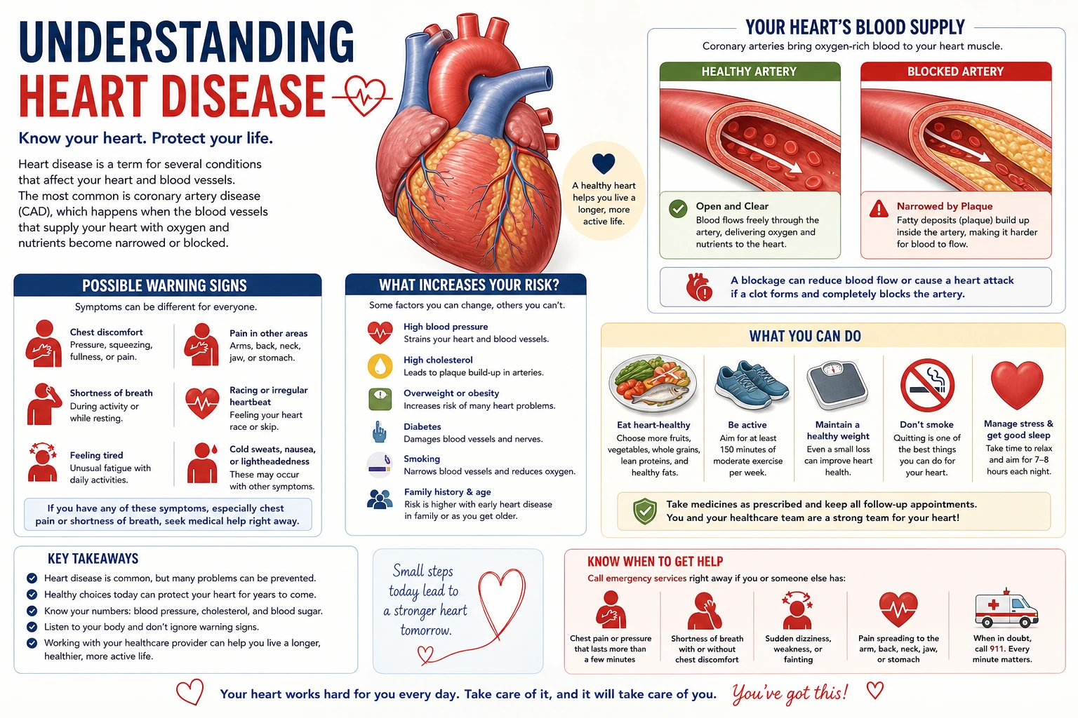 Patient Education
Move to a patient-facing page for eye-care, visit-preparation, or recovery explainers.