
Skin Layers - Cross-Section
Layered skin anatomy with epidermis, dermis, subcutis, adnexal structures, and superficial vessels.
anatomy
Draft dermatology visuals for skin-layer teaching, lesion morphology, melanoma depth explanation, wound-care communication, and biopsy or Mohs overview content. Use this page for explanation and review, not for diagnostic sign-off.
Free to try ·
Your diagram will appear here
Describe what you need and click Generate
Skin diagrams for lesion morphology, melanoma staging, wound care, and dermatologic procedure teaching
Layered skin anatomy with epidermis, dermis, subcutis, adnexal structures, and superficial vessels.
Side-by-side comparison of eczema, psoriasis, and acne using standard lesion morphology.
Reference panel for primary lesion classification with consistent labels and scale.
Cutaneous melanoma diagram with Breslow depth and Clark level landmarks across skin layers.
Four-panel wound healing sequence from clot formation to remodeling and scar maturation.
Procedure panel comparing biopsy depth, specimen type, and Mohs staged margin control.
A useful dermatology figure shows where the process sits in the skin, how the lesion is described, and how the pattern distributes across the body or within the specimen. That clarity helps with teaching, counseling, and biopsy explanation.
Use it for skin-layer teaching, lesion-description review, wound-healing communication, and high-level procedural explanation rather than for pathology slide interpretation or final diagnosis.
For resident teaching, keep morphology and depth explicit. For patient materials, reduce label density and use sequence or recovery cues. If the figure needs heavy histology comparison, the Pathology Diagram Generator is usually the better companion.
Start with the lesion type, body location, and depth relationship. Then say whether the image is for clinic explanation, trainee review, or procedure counseling.
Build skin layer, lesion morphology, melanoma depth, wound care, and procedure figures for rounds, resident teaching, and patient counseling.
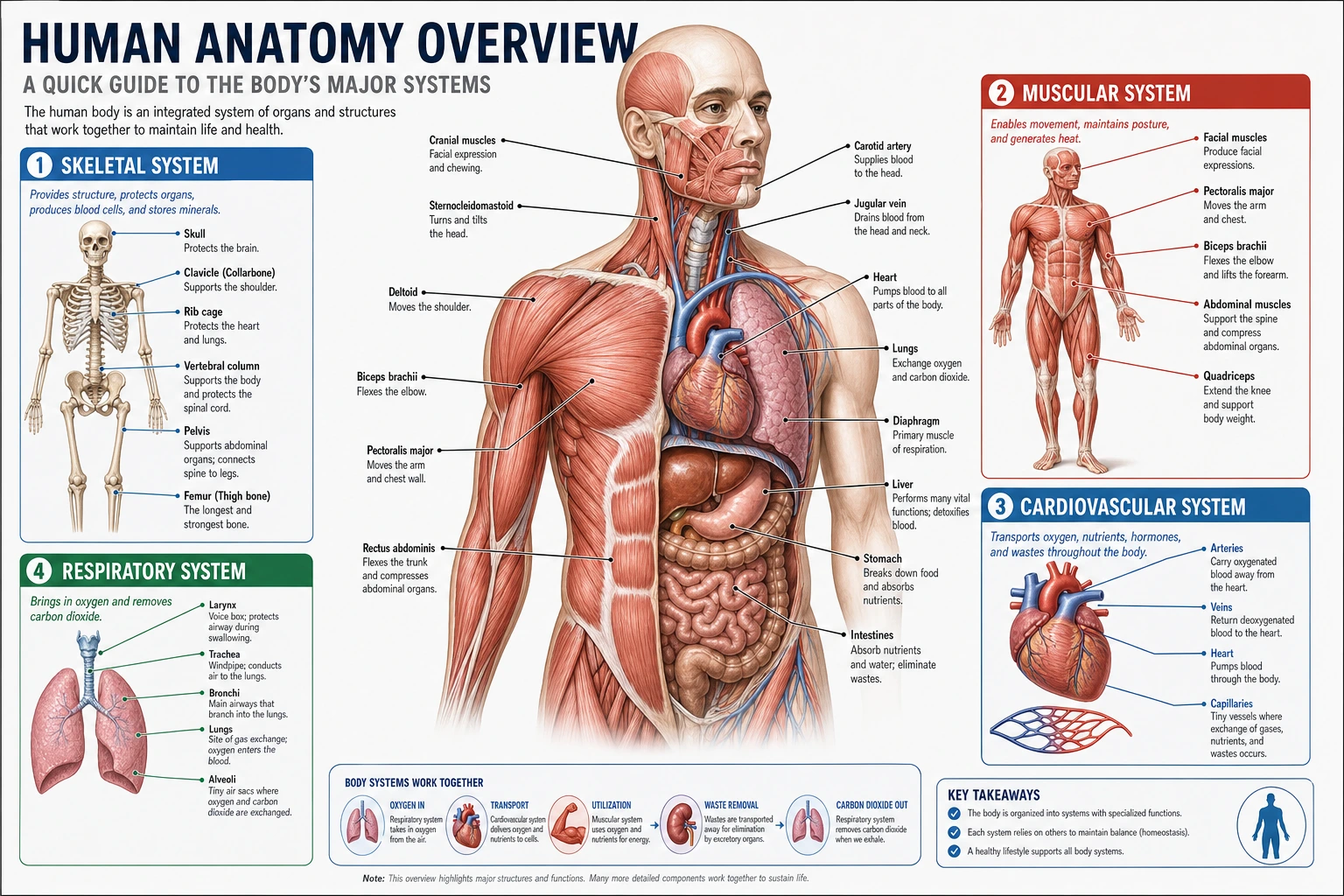 Anatomy
Use broader anatomy plates when a skin figure needs surrounding nerves, vessels, or regional landmarks.
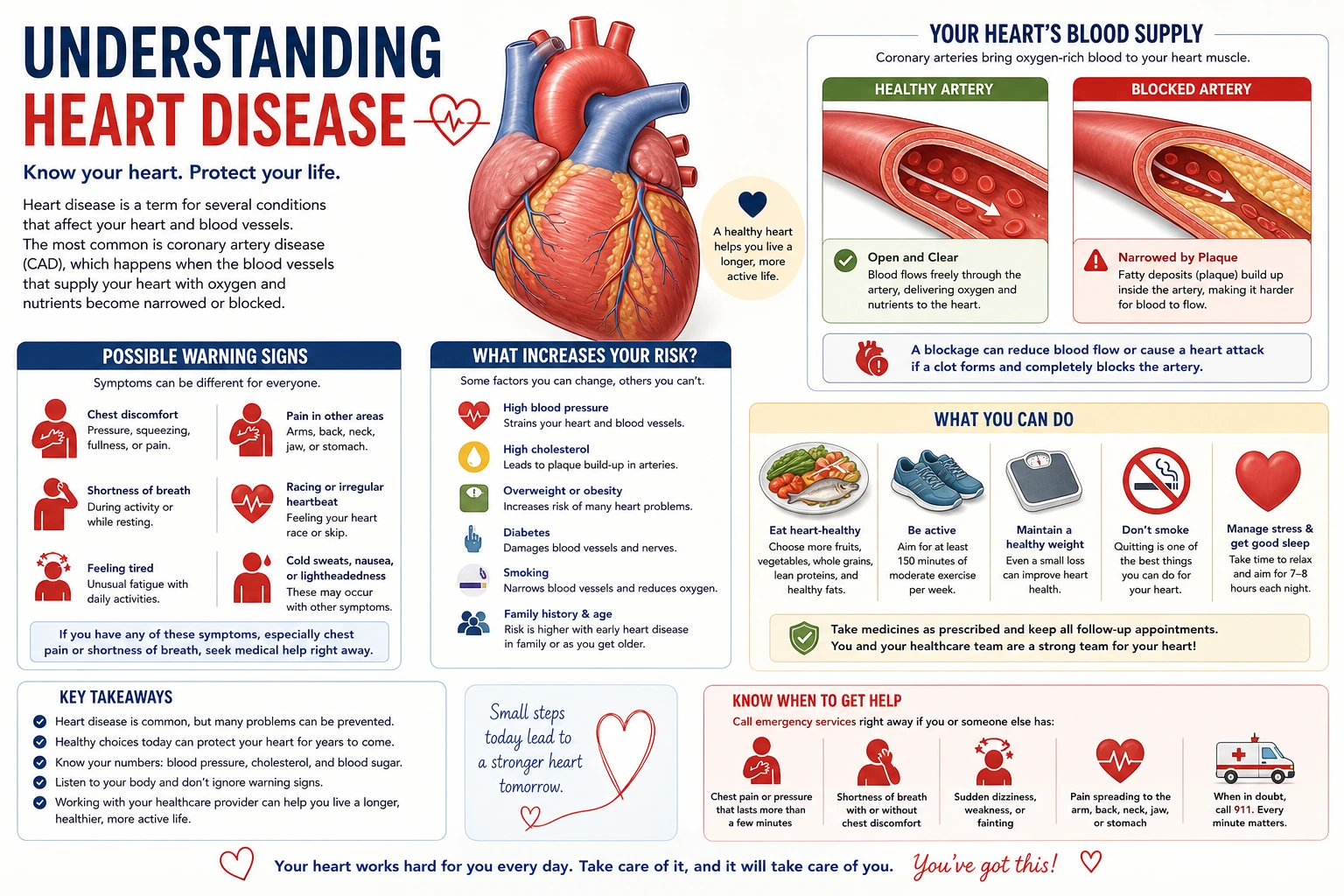 Patient Education
Create simpler skin care, biopsy aftercare, and treatment explanation visuals for handouts.
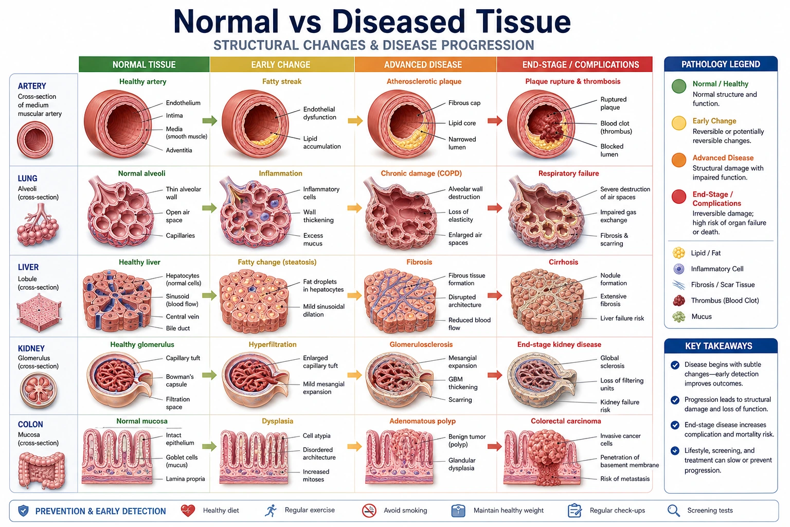 Clinical
Pair clinical skin diagrams with histology-focused disease comparisons and tissue pattern panels.